
Introduction to Protein Dephosphorylation and Cellular Signaling
Phosphorylation and dephosphorylation are among the most fundamental biochemical processes that regulate cellular physiology. Within living organisms, phosphate ester bonds play essential roles in energy metabolism, nucleic acid synthesis, intracellular communication, and enzymatic regulation. The reversible addition and removal of phosphate groups from proteins acts as a molecular switch that controls the activity, localization, stability, and interactions of thousands of cellular proteins. Through this dynamic mechanism, cells are able to rapidly respond to environmental changes, hormonal stimulation, stress conditions, nutrient availability, and developmental signals.
Protein phosphorylation primarily occurs on the hydroxyl-containing amino acids serine, threonine, and tyrosine. The phosphorylation process is catalyzed by protein kinases, whereas the removal of phosphate groups is mediated by protein phosphatases. Together, these enzymes form highly coordinated signaling networks that regulate virtually every aspect of eukaryotic cell biology, including gene transcription, metabolism, cell-cycle progression, apoptosis, neuronal communication, immune activation, cytoskeletal organization, and differentiation.
Although protein kinases have historically received considerable scientific attention, protein phosphatases are equally important regulators of signal transduction. Rather than acting merely as passive “off switches,” phosphatases function as highly specific and tightly controlled enzymes capable of integrating multiple signaling pathways. The balance between kinase and phosphatase activity ultimately determines the phosphorylation state of intracellular proteins and therefore controls the functional output of signaling cascades.
Protein phosphatases are generally classified into two major groups according to substrate specificity. Serine/threonine protein phosphatases hydrolyze phosphoserine and phosphothreonine residues, whereas protein tyrosine phosphatases specifically remove phosphate groups from phosphotyrosine residues. A specialized subgroup known as dual-specificity phosphatases possesses the ability to dephosphorylate both serine/threonine and tyrosine residues, allowing these enzymes to participate in more complex regulatory pathways.
Recent advances in structural biology, particularly X-ray crystallography, have significantly improved the understanding of phosphatase architecture, catalytic mechanisms, substrate recognition, and regulatory control. Structural analyses have demonstrated that serine/threonine phosphatases and tyrosine phosphatases employ fundamentally different catalytic strategies despite performing the same overall biochemical reaction of phosphoester hydrolysis. This divergence contrasts sharply with protein kinases, which generally share a conserved catalytic fold and mechanistic framework.

Structural Organization of Serine/Threonine Protein Phosphatases
Serine/Threonine Phosphatase Families
Serine/threonine protein phosphatases represent a large family of metalloenzymes that participate in numerous essential cellular functions. These enzymes regulate metabolic pathways, cytoskeletal dynamics, cell-cycle checkpoints, stress responses, transcriptional control, and intracellular communication. Their activities are precisely controlled through association with regulatory proteins, post-translational modifications, and subcellular localization mechanisms.
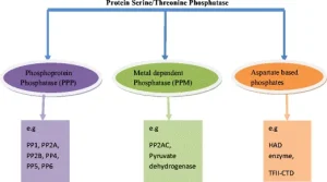
Comparative amino acid sequence analysis revealed the existence of two major serine/threonine phosphatase gene families. One family includes protein phosphatase 1 (PP1), protein phosphatase 2A (PP2A), and calcineurin (PP2B), all of which share significant sequence homology within their catalytic domains. In contrast, protein phosphatase 2C (PP2C) belongs to a structurally distinct family that evolved independently and possesses different catalytic properties.
Each phosphatase family fulfills specialized biological roles:
- Calcineurin (PP2B) is a calcium/calmodulin-dependent phosphatase involved in T-cell activation and immune signaling.
- PP1 regulates glycogen metabolism, muscle contraction, protein synthesis, and neuronal signaling.
- PP2A controls cell-cycle progression, DNA replication, apoptosis, and transcriptional regulation.
- PP2C participates in stress adaptation and osmotic regulation, particularly in yeast and plant systems.
The structural elucidation of PP1 and calcineurin provided major insights into the molecular basis of serine/threonine dephosphorylation and demonstrated that these enzymes share a conserved catalytic architecture centered around a metal-dependent active site.
Conserved Structural Core of Serine/Threonine Phosphatases
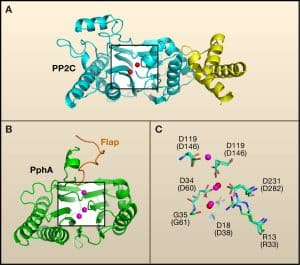
Crystal structures of PP1 and calcineurin revealed a highly conserved catalytic scaffold composed of alternating beta strands and alpha helices that form a central β-α-β-α-β structural motif. At the core of this architecture lies a dinuclear metal center that is essential for catalytic activity and structural stability.
The metal-binding site is coordinated by highly conserved amino acid residues, including:
- Three histidine residues
- Two aspartic acid residues
- One asparagine residue
These coordinating residues are positioned primarily within flexible loops connecting secondary structural elements. The arrangement of these residues creates an optimal environment for metal ion binding and catalytic water activation.
Interestingly, different divalent metal ions such as iron, zinc, and manganese can occupy the catalytic center. This flexibility suggests that enzymatic activity may be influenced not only by protein structure but also by the identity and oxidation state of the bound metals. Such metal-dependent regulation may allow cells to modulate phosphatase activity in response to metabolic conditions or oxidative stress.
Structural comparisons further demonstrated that serine/threonine phosphatases belong to a broader superfamily of metallo-phosphoesterases that includes bacterial exonucleases, RNA-processing enzymes, and purple acid phosphatases. This observation indicates that evolution repeatedly utilized a highly efficient catalytic framework for phosphate ester hydrolysis across multiple biological systems.
Structural Features of Protein Tyrosine Phosphatases
Functional Diversity of Protein Tyrosine Phosphatases
Protein tyrosine phosphatases are critical regulators of signal transduction pathways that control cellular growth, proliferation, differentiation, migration, metabolism, and immune responses. Depending on cellular context, PTPs may either positively or negatively regulate signaling pathways.
Examples include:
- SHP-2, which promotes Ras pathway activation
- SHP-1, which suppresses immune receptor signaling
- CDC25 phosphatases, which activate cyclin-dependent kinases during cell-cycle progression
- MAP kinase phosphatases (MKPs), which terminate MAPK signaling pathways
Unlike serine/threonine phosphatases, PTPs do not require metal ions for catalysis. Instead, they utilize a catalytic cysteine residue positioned within a highly conserved active-site signature motif.
This catalytic sequence forms the structural foundation of the phosphotyrosine-binding pocket.
Catalytic Domain Architecture of PTPs
The catalytic domains of PTPs are generally composed of approximately 200–300 amino acids and adopt a compact α/β fold. Four parallel beta strands form a central core that is surrounded by alpha helices. The active-site loop containing the HCXXGXXRS(T) motif is positioned between a beta strand and an alpha helix.
Several residues within this motif are essential for catalysis:
- The cysteine residue acts as the nucleophile
- The arginine residue stabilizes phosphate binding
- Backbone amides help coordinate substrate phosphates
Approximately 30–40 residues upstream of the catalytic cysteine lies an essential aspartic acid residue located on a flexible loop. This loop undergoes large conformational rearrangements during substrate binding and catalysis, positioning the aspartic acid for proton transfer reactions.
Despite very limited sequence identity between classical PTPs and dual-specificity phosphatases, X-ray structures demonstrated remarkable conservation of the overall catalytic fold. This structural similarity highlights the evolutionary importance of the PTP catalytic architecture.

Structural Determinants of Substrate Specificity
Active-Site Depth and Dual Specificity
One of the most important structural determinants of substrate specificity among phosphatases is the geometry of the active-site pocket.
Classical tyrosine phosphatases possess deep active-site clefts approximately 9 Å in depth. This configuration allows phosphotyrosine residues, which contain bulky aromatic rings, to access the catalytic cysteine positioned at the base of the pocket.
In contrast, dual-specificity phosphatases possess shallower active-site pockets measuring approximately 6 Å deep. The reduced depth enables these enzymes to accommodate both phosphotyrosine and the smaller phosphoserine/phosphothreonine residues.
Structural studies identified a flexible loop located between the first alpha helix and first beta strand as a major determinant of pocket depth and substrate recognition. Variations in the length and composition of this loop dramatically alter catalytic specificity and substrate accessibility.
This structural adaptation provides an elegant molecular explanation for how closely related phosphatases evolved distinct substrate preferences while maintaining a conserved catalytic core.
Catalytic Mechanisms of Protein Phosphatases
Catalysis by Serine/Threonine Protein Phosphatases
Serine/threonine phosphatases utilize a metal-dependent hydrolytic mechanism that does not involve formation of a covalent phosphoenzyme intermediate.
The catalytic process is thought to proceed through direct nucleophilic attack of an activated water molecule on the phosphorus atom of the substrate phosphate group. The water molecule is bridged between the two catalytic metal ions, which help lower its pKa and increase nucleophilicity.
The proposed mechanism includes:
- Activation of a bridging water molecule by the dinuclear metal center
- Nucleophilic attack on the substrate phosphate
- Cleavage of the phosphoester bond
- Release of inorganic phosphate and dephosphorylated substrate
A nearby histidine residue likely functions as a general acid by protonating the leaving group oxygen during catalysis. Adjacent acidic residues may further stabilize transition states and orient catalytic residues for efficient proton transfer.
Although substantial evidence supports this catalytic model, many mechanistic details remain under investigation, particularly regarding transition-state stabilization and substrate recognition dynamics.

Catalysis by Protein Tyrosine Phosphatases
In contrast to serine/threonine phosphatases, protein tyrosine phosphatases employ a covalent catalytic mechanism involving formation of a phosphocysteine intermediate.
The reaction proceeds through two distinct chemical steps:
Step 1: Formation of the Phosphoenzyme Intermediate
The catalytic cysteine exists as a highly reactive thiolate anion. During catalysis, this thiolate attacks the phosphorus atom of the substrate phosphate group.
The substrate phosphate is precisely positioned within the active site through multiple hydrogen bonds involving:
- The catalytic arginine residue
- Backbone amide groups
- Conserved active-site residues
At the same time, the catalytic aspartic acid donates a proton to the leaving group oxygen, facilitating cleavage of the phosphoester bond.
This reaction generates:
- A covalent phosphocysteine intermediate
- A dephosphorylated protein substrate
Step 2: Hydrolysis of the Intermediate
The phosphoenzyme intermediate must then be hydrolyzed to regenerate the active enzyme.
A water molecule attacks the phosphocysteine bond, releasing inorganic phosphate. The catalytic aspartic acid likely functions as a general base during this step by activating the hydrolytic water molecule.
This two-step mechanism provides exceptional catalytic efficiency and specificity while allowing tight regulation of enzymatic activity.
Conformational Dynamics During Catalysis
Structural studies demonstrated that catalysis by PTPs involves major conformational rearrangements.
A flexible loop containing the catalytic aspartic acid shifts dramatically upon substrate binding. In the inactive “open” conformation, the aspartic acid is positioned far from the active site. Upon substrate engagement, the loop closes over the active site and properly positions the aspartic acid for catalysis.
This dynamic movement:
- Enhances substrate specificity
- Protects reaction intermediates
- Stabilizes transition states
- Coordinates proton transfer events
Such conformational flexibility illustrates the sophisticated regulatory mechanisms that govern phosphatase function at the molecular level.

Receptor-Like Protein Tyrosine Phosphatases
Structural Regulation Through Dimerization
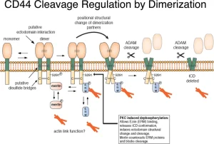
Receptor-like protein tyrosine phosphatases contain extracellular domains, transmembrane regions, and intracellular catalytic domains. Many possess two intracellular phosphatase domains, although only one is typically catalytically active.
Structural studies revealed that receptor-like PTPs may be regulated through dimerization mechanisms. In some cases, an amino-terminal helix-turn-helix segment from one phosphatase molecule inserts directly into the active site of another molecule within a dimeric complex.
This interaction effectively blocks substrate access and inhibits catalytic activity. Such findings suggest that receptor dimerization may function as an important physiological mechanism for controlling phosphatase signaling at the cell surface.
Evolutionary Relationships Among Phosphatases
Convergent and Divergent Evolution
Although serine/threonine phosphatases and tyrosine phosphatases catalyze the same overall biochemical reaction, they evolved fundamentally different structural frameworks and catalytic strategies.
Serine/threonine phosphatases rely on:
- Metal-dependent catalysis
- Activated water nucleophiles
- Dinuclear metal centers
Tyrosine phosphatases utilize:
- Catalytic cysteine residues
- Covalent phosphoenzyme intermediates
- Acid-base catalysis
Interestingly, low molecular weight phosphatases independently evolved catalytic motifs resembling those of PTPs despite possessing entirely unrelated protein folds. This phenomenon represents an excellent example of convergent evolution, where similar biochemical solutions arise independently in different protein families.
Biological Importance of Protein Dephosphorylation
Protein phosphatases regulate virtually every major cellular process. Their biological functions include:
- Immune system activation
- Hormonal signaling
- Synaptic plasticity
- Cell-cycle progression
- DNA replication
- Stress responses
- Apoptosis
- Metabolic regulation
- Developmental signaling
Because phosphatases function within tightly interconnected signaling networks, dysregulation of phosphatase activity contributes to numerous diseases, including:
- Cancer
- Diabetes
- Neurodegenerative disorders
- Autoimmune diseases
- Cardiovascular disease
Understanding phosphatase structure and catalytic function therefore has major implications for therapeutic drug development.

Future Perspectives in Protein Phosphatase Research
Recent advances in structural biology, enzymology, and molecular signaling have transformed the understanding of protein phosphatases. Detailed structural characterization has clarified catalytic mechanisms, substrate specificity determinants, conformational dynamics, and regulatory interactions.
Future research directions include:
- Development of highly selective phosphatase inhibitors
- Identification of phosphatase regulatory complexes
- Investigation of phosphatase signaling networks
- Structural analysis of transient catalytic intermediates
- Exploration of phosphatase roles in disease progression
Because phosphatases control essential signaling pathways, they represent highly promising therapeutic targets. Continued investigation into the molecular mechanisms governing protein dephosphorylation will provide deeper insight into cellular regulation and may lead to novel strategies for treating human disease.